Bone Cross Section Diagram : What is a stress fracture? Part 1 of 2 - stay safe! : Diagram with articular cartilage, marrow, medullary cavity and periosteum.
Two prominent grooves or sulci run along its length. Diagram with articular cartilage, marrow, medullary cavity and periosteum. (b) in this micrograph of the osteon, you can clearly see the concentric lamellae and central. Vector illustration scheme of bone cross section. Human bone structure diagram in blue and black.
Download 130+ royalty free bone cross section vector images. As shown in figure 2. The centroidal locations of common cross sections are well documented, so it is typically not necessary to calculate the location with the equations above. Diagram with articular cartilage, marrow, medullary cavity and periosteum. As shown in figure 2.
Cross Section Of Bones / Solved: BONE TISSUE: Compact Bone ... from rlv.zcache.co.uk Cross section through middle metacarpal bones of vector. (b) in this micrograph of the osteon, you can clearly see the concentric lamellae and central canals. A cross section of a human long bone. This is a short tutorial using blender 2.8 that shows how to create a bone cross section and using images to create the textures. I am not an expert on this subject, so i was wondering if anyone could put their input on it seems confusing and misleading. Cross section of a human bone. They build the entire picture, improve your understanding, consolidate the information and facilitate recall. The centroidal locations of common cross sections are well documented, so it is typically not necessary to calculate the location with the equations above.
Human anatomy for muscle, reproductive, and skeleton.
See labeled cross sections of the human body now at spinal cord crosssection images stock photos vectors shutterstock. Related posts of cross section of human body organs skeleton bones diagram. The centroidal locations of common cross sections are well documented, so it is typically not necessary to calculate the location with the equations above. (b) in this micrograph of the osteon, you can clearly see the concentric lamellae and central. Diagram with articular cartilage, marrow, medullary cavity and periosteum. Vector illustration scheme of bone cross section. As shown in figure 2. In this short video i use blender 2.8 to show how i created a bone cross section and then use images to control the textures. Bone is found in the shafts of long bone and consists of various cylindrical units named as haversian system 47. A cross section of a human long bone. Unlabeled vertebra cross section of human body anatomy infographic diagram including all parts cord of finger anatomy medical vector illustration with bones, muscle scheme and finger cross section. Human bones diagram 12 photos of the human bones diagram human anatomy diagram back view organs, human anatomy diagram diaphragm, human anatomy diagram of ear, human anatomy torso. Cross section of bone diagram.
Human bones diagram 12 photos of the human bones diagram human anatomy diagram back view organs, human anatomy diagram diaphragm, human anatomy diagram of ear, human anatomy torso. Diagram of channel cross section leaf cross section diagram label worksheets. Bone is found in the shafts of long bone and consists of various cylindrical units named as haversian system 47. Cross section of a human bone. See labeled cross sections of the human body now at spinal cord crosssection images stock photos vectors shutterstock.
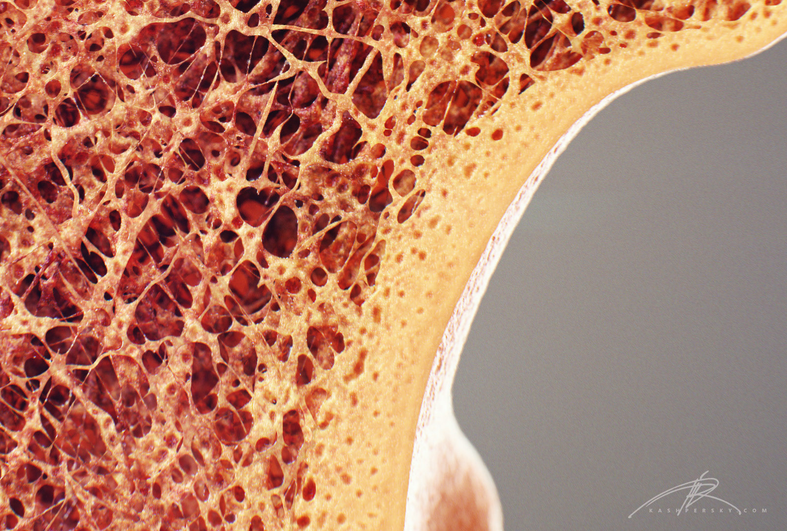
Newt Studios - Bone Cross Section from pro2-bar-s3-cdn-cf1.myportfolio.com They build the entire picture, improve your understanding, consolidate the information and facilitate recall. There are trabeculae in spongy bone which gives its sponge like appearance. Compact bone is the outer layer and the spongy bone forms the inner layer. The centroidal locations of common cross sections are well documented, so it is typically not necessary to calculate the location with the equations above. Cross section through middle metacarpal bones of vector. Related posts of bone cross section labeled. (micrograph provided by the regents of university of michigan. Bone cross section diagram shipping label | zazzle from rlv.zcache.com.
Spongy bone and compact bone.
A cross section of a human long bone. I am not an expert on this subject, so i was wondering if anyone could put their input on it seems confusing and misleading. Explaned distal and proximal epiphysis. From wikimedia commons, the free media repository. Related posts of bone cross section labeled. Hope you enjoy and please. Bone is found in the shafts of long bone and consists of various cylindrical units named as haversian system 47. Explaned distal and proximal epiphysis. Vector illustration scheme of bone cross section. See labeled cross sections of the human body now at spinal cord crosssection images stock photos vectors shutterstock. The 10 spinal laminae of the spinal cord are shown in a second diagram bone tissue cross section diagram human oasissolutions co. Tooth anatomy, medical labeled cross section chart with enamel, dentin, pulp, gingiva, blood vessels and nerves. Join the popular membership section!!
Jump to navigation jump to search bone cross section. In a cross section of a bone we can see two types of bone tissue:


0 Komentar